41 microscope images with labels
Polarizing Microscope Image Gallery - Leica Microsystems The position of the optical axis can be clearly determined with circular polarization. Right: Conoscopic image of the same calcite sample with linear polarized light. The calcite section is perpendicular to the optical axis. Images recorded with a DM4 P microscope using transmitted light, conoscopy, 63x N Plan objective, and polarizers. Microscope Labeling - The Biology Corner Students label the parts of the microscope in this photo of a basic laboratory light microscope. Can be used for practice or as a quiz. ... The type of microscope used in most science classes is the _____ microscope. 18. You should carry the microscope by the _____ and the _____. 19. The objectives are attached to what part of the microscope ...
Amazing 27 Things Under The Microscope With Diagrams The tail is transparent and thus is difficult to detect under a low-power microscope. 23. Spirogyra under the microscope. Spirogyra is a green alga found mostly in freshwater in the form of green clumps. Spirogyra is unicellular, but because it clumps together, it can be seen in the pond even with our naked eyes.

Microscope images with labels
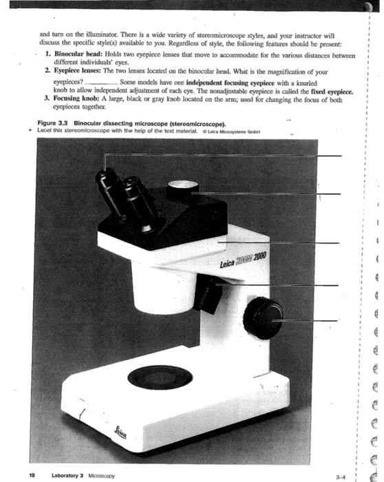
Microscope Labeling Game - PurposeGames.com About this Quiz. This is an online quiz called Microscope Labeling Game. There is a printable worksheet available for download here so you can take the quiz with pen and paper. This quiz has tags. Click on the tags below to find other quizzes on the same subject. Science. Labeling a Microscope Free Worksheet Pack More like this. These parts of a microscope printables include word searches, crossword puzzles, and vocabulary worksheets. Learn about animal and plant cells with these printable worksheets. Budding biologists, here's a great cheat-sheet to help you memorize and compare the structures of both animal cells and plant cells. rsscience.com › stereo-microscopeParts of Stereo Microscope (Dissecting microscope) - Rs' Science Unlike a compound microscope that offers a flat image, stereo microscopes give the viewer a 3-dimensional image that you can see the texture of a larger specimen. [In this image] Examples of Stereo & Dissecting microscopes. Major microscope brands (Zeiss, Olympus, Nikon, Amscope, Omano, Leica …) all produce stereomicroscopes.
Microscope images with labels. Compound Microscope Parts, Functions, and Labeled Diagram Compound Microscope Definitions for Labels. Eyepiece (ocular lens) with or without Pointer: The part that is looked through at the top of the compound microscope. Eyepieces typically have a magnification between 5x & 30x. Monocular or Binocular Head: Structural support that holds & connects the eyepieces to the objective lenses. › products › microscopeLAS X Industry Microscope software for Industry | Products ... Create a single sharp image by capturing a stack of images at different focus positions and combining them automatically into an Extended Depth of Focus (EDOF) image. LAS X Extended Depth of Field: Create sharp 2D images from several partially in-focus images. In connection with the 3D Surface Viewer, creation of 3D images is also possible. Parts of the Microscope with Labeling (also Free Printouts) Microscopes are specially created to magnify the image of the subject being studied. This exercise is created to be used in homes and schools. the microscope layout, including the blank and answered versions are available as pdf downloads. Click to Download : Label the Parts of the Microscope (A4) PDF print version. Microscope, Microscope Parts, Labeled Diagram, and Functions Revolving Nosepiece or Turret: Turret is the part of the microscope that holds two or multiple objective lenses and helps to rotate objective lenses and also helps to easily change power. Objective Lenses: Three are 3 or 4 objective lenses on a microscope. The objective lenses almost always consist of 4x, 10x, 40x and 100x powers. The most common eyepiece lens is 10x and when it coupled with ...
Microscope picture label Flashcards | Quizlet Start studying Microscope picture label. Learn vocabulary, terms, and more with flashcards, games, and other study tools. PDF Label parts of the Microscope Label parts of the Microscope: . Created Date: 20150715115425Z Explanation and Labelled Images - New York Microscope Company More than one method exists, and the most common ones include labelling with fluorescent stains, expression of a fluorescent protein, or taking advantage of the intrinsic fluorescence (autofluorescence) of a sample. Fluorescence microscopy is a common and powerful tool in the life sciences. Epifluorescence microscopy Microscope Labeling - The Biology Corner Microscope Labeling. Shannan Muskopf May 31, 2018. This simple worksheet pairs with a lesson on the light microscope, where beginning biology students learn the parts of the light microscope and the steps needed to focus a slide under high power. The labeling worksheet could be used as a quiz or as part of direct instruction where students label the microscope as you go over what each part is used for.
16 Parts of a Compound Microscope: Diagrams and Video It's actually not a toy microscope, it's a functional microscope that produces great images for the price. I bought it for less than $100 dollars but you can check the current price on Amazon. 1. Head (Body) The head, also referred to as the body of the microscope, is a structural component that contains the optical parts of the microscope. Compound Microscope Parts - Labeled Diagram and their Functions - Rs ... There are two major optical lens parts of a microscope: Eyepiece (10x) and Objective lenses (4x, 10x, 40x, 100x). Total magnification power is calculated by multiplying the magnification of the eyepiece and objective lens. The illuminator provides a source of light. The light is focused by the condenser and passing through the specimen placed ... Microscope Labeled Pictures, Images and Stock Photos Browse 48 microscope labeled stock photos and images available, or start a new search to explore more stock photos and images. Newest results Fluorescent Imaging immunofluorescence of cancer cells growing in 2D with nuclei in blue, cytoplasm in red and DNA damage foci in green microscope labeled stock pictures, royalty-free photos & images Microscope Parts, Function, & Labeled Diagram - slidingmotion Condenser. The condenser is to focus the light, which passes from the microscopic illuminator to the specimen. This condenser is located just below the diaphragm. This diaphragm is one of the important parts of the compound microscope which will help to get an accurate and sharp image. The condenser has a magnification power of 400X and above.
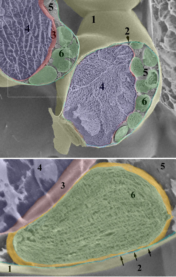
Leaf chloroplast
Electron Microscopy Images - Dartmouth Transmission electron microscope image of a thin section cut through the bronchiolar epithelium of the lung (mouse), which consists of ciliated cells and non-ciliated cells. Image shows the ciliary microtubules in transverse and oblique section. In the cell apex are the basal bodies that are the anchoring sites for the cilia.

MICROBIOLOGY SLIDE SPECIMENS
› products › microscopeMicroscope Objective Lens | Products | Leica Microsystems The objective lens is a critical part of the microscope optics. The microscope objective is positioned near the sample, specimen, or object being observed. It has a very important role in imaging, as it forms the first magnified image of the sample. The numerical aperture (NA) of the objective indicates its ability to gather light and largely determines the microscope’s resolution, the ...

35 Label Of Compound Microscope - Labels For Your Ideas
Skin Images Labeled | Virtual Anatomy Lab VAL - ncccval Skin Images Labeled | Virtual Anatomy Lab VAL ... Connective Tissue Images Unlabeled. Microscope. Microscope Images Labeled. Microscope Images Unlabeled. Mitosis. Mitosis Images Labeled. Mitosis Images Unlabeled. Skin. Skin Images Labeled. Skin Images Unlabeled. Skeletal system. Skeletal Images Labeled. Skeletal Images Unlabeled.
31 Label The Indicated Parts Of The Microscope - Labels Database 2020
300+ Free Microscope & Laboratory Images - Pixabay Find your perfect microscope image. Free pictures to download and use in your next project. 189 37. analysis biochemistry. 335 71. analysis biochemistry. 334 96. microscope slide. 725 186.

microscope labeled 5406509 orig - Top Label Maker
Labeling the Parts of the Microscope Labeling the Parts of the Microscope. This activity has been designed for use in homes and schools. Each microscope layout (both blank and the version with answers) are available as PDF downloads. You can view a more in-depth review of each part of the microscope here.
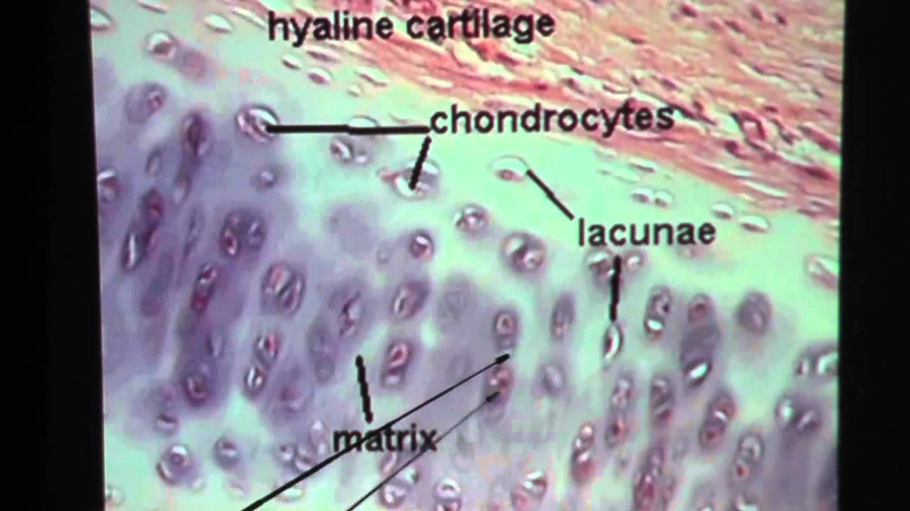
Hyaline Cartilage Connective Tissue - YouTube
Compound Microscope - Diagram (Parts labelled), Principle and Uses Image : Labeled Diagram of compound microscope parts. See: Labeled Diagram showing differences between compound and simple microscope parts Structural Components. The three structural components include. 1. Head. This is the upper part of the microscope that houses the optical parts. 2. Arm . This part connects the head with the base and provides stability to the microscope.

Labeling The Microscope
Microscope Drawing And Label - Painting Valley All the best Microscope Drawing And Label 33+ collected on this page. Feel free to explore, study and enjoy paintings with PaintingValley.com. ... All rights to paintings and other images found on PaintingValley.com are owned by their respective owners (authors, artists), and the Administration of the website doesn't bear responsibility for ...
33 Label Of Compound Microscope - Labels Database 2020
› microscopy › intZEISS Axiocam Microscope Cameras for Science and Research For easy documentation, some digital cameras can record images either completely without a computer, or through a connected PC, laptop or iPad running the ZEISS Labscope software. All our microscope cameras are fully supported in our ZEN software environment, offering fast live image display and easy-to-use user interface.
Search in gallery
Microscope Types (with labeled diagrams) and Functions A compound microscope: Is used to view samples that are not visible to the naked eye. Uses two types of lenses - Objective and ocular lenses. Has a higher level of magnification - Typically up to 2000x. Is used in hospitals and forensic labs by scientists, biologists and researchers to study micro organisms. Compound microscope labeled diagram.

Labeling a Compound Microscope
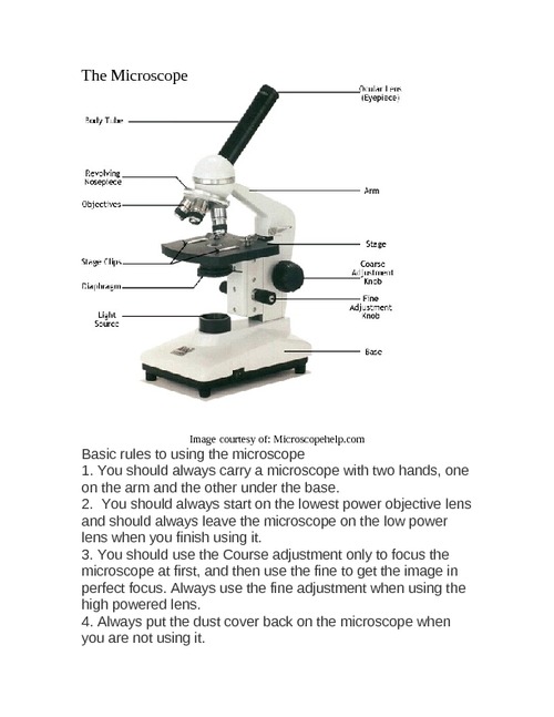
Microscope Parts and Functions Eyepiece: The lens the viewer looks through to see the specimen. The eyepiece usually contains a 10X or 15X power lens. Diopter Adjustment: Useful as a means to change focus on one eyepiece so as to correct for any difference in vision between your two eyes. Body tube (Head): The body tube connects the eyepiece to the objective lenses. Arm: The arm connects the body tube to the base of the ...

Best Top Desktop Wallpapers: Electron microscope images
Label the microscope — Science Learning Hub All microscopes share features in common. In this interactive, you can label the different parts of a microscope. Use this with the Microscope parts activity to help students identify and label the main parts of a microscope and then describe their functions. Drag and drop the text labels onto the microscope diagram. If you want to redo an answer, click on the box and the answer will go back to the top so you can move it to another box.
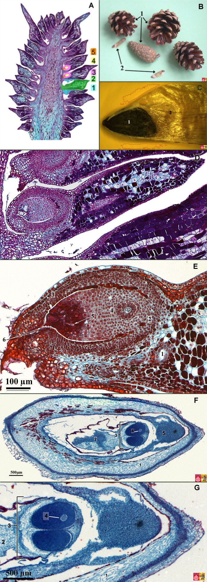
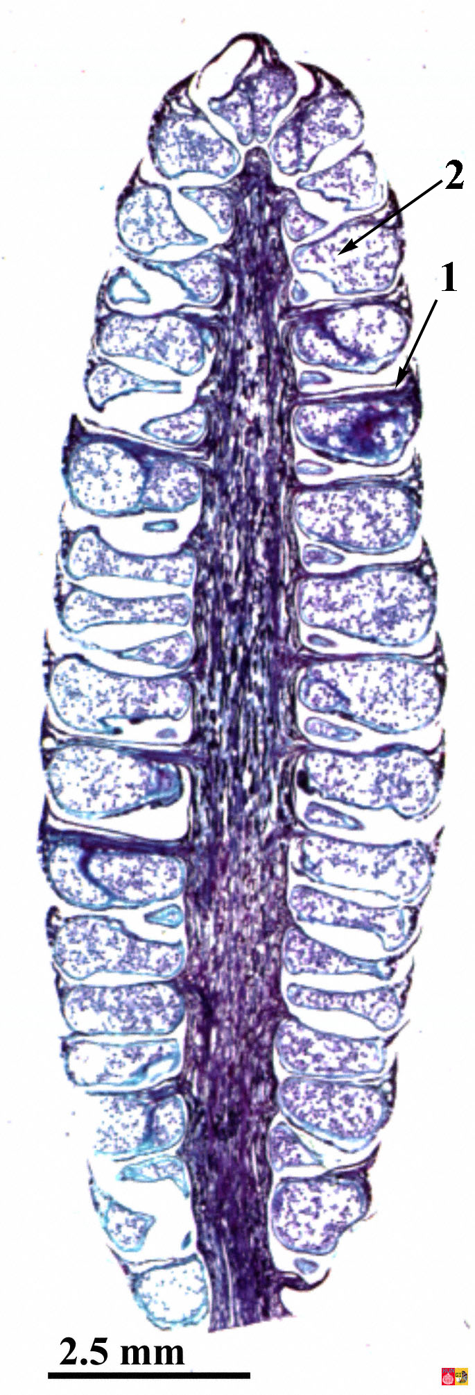
Life Cycle of the Pine Tree (Gymnosperm)
Histology and Microscope Slide Labels Microscope Slide Labels. These specialty Microscope Slide Labels and matching End Labels are available in standard (thin) or pathology (tissue high) thickness, and square or round corner (RC). Permanent adhesive holds labels in place during use and long-term storage. Sheet Form Size is 5¼" x 8". Prices are per thousand labels. Slide Label

microscope labeled microscope worksheet labeling sc 1 st template entrancing labelling - Top ...
Microscope With Labels clip art | Microscope parts, Scientific method ... Jul 3, 2012 - Download Clker's Microscope With Labels clip art and related images now. Multiple sizes and related images are all free on Clker.com.

32 Picture Of Microscope With Label - Labels For You
› books › NBK26880Looking at the Structure of Cells in the Microscope A typical animal cell is 10–20 μm in diameter, which is about one-fifth the size of the smallest particle visible to the naked eye. It was not until good light microscopes became available in the early part of the nineteenth century that all plant and animal tissues were discovered to be aggregates of individual cells.

Endocrine Pancreas
› dinocaptureDinoCapture 2.0: Microscope Imaging Software | Dino-Lite Dino-Lite USB microscope cameras include DinoCapture 2.0, the powerful yet easy to use microscope imaging software for Windows. DinoCapture is a professional microscope imaging software that was made for users of all levels, including basic features from image viewing and capture, measurement with calibration, to advanced features such as geotags and edge detection.
Zoeken in galerij
en.wikipedia.org › wiki › Electron_microscopeElectron microscope - Wikipedia An electron microscope is a microscope that uses a beam of accelerated electrons as a source of illumination. As the wavelength of an electron can be up to 100,000 times shorter than that of visible light photons , electron microscopes have a higher resolving power than light microscopes and can reveal the structure of smaller objects.
Post a Comment for "41 microscope images with labels"