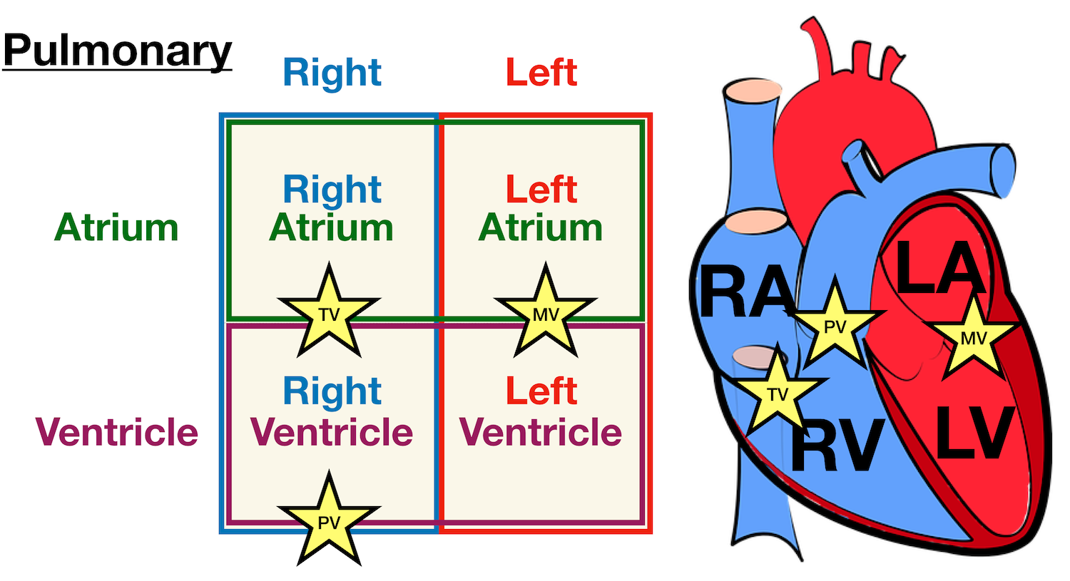
40 heart structure and labels
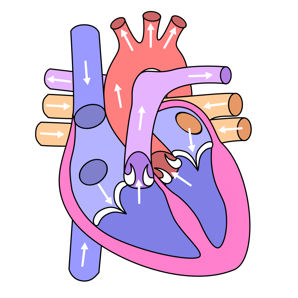
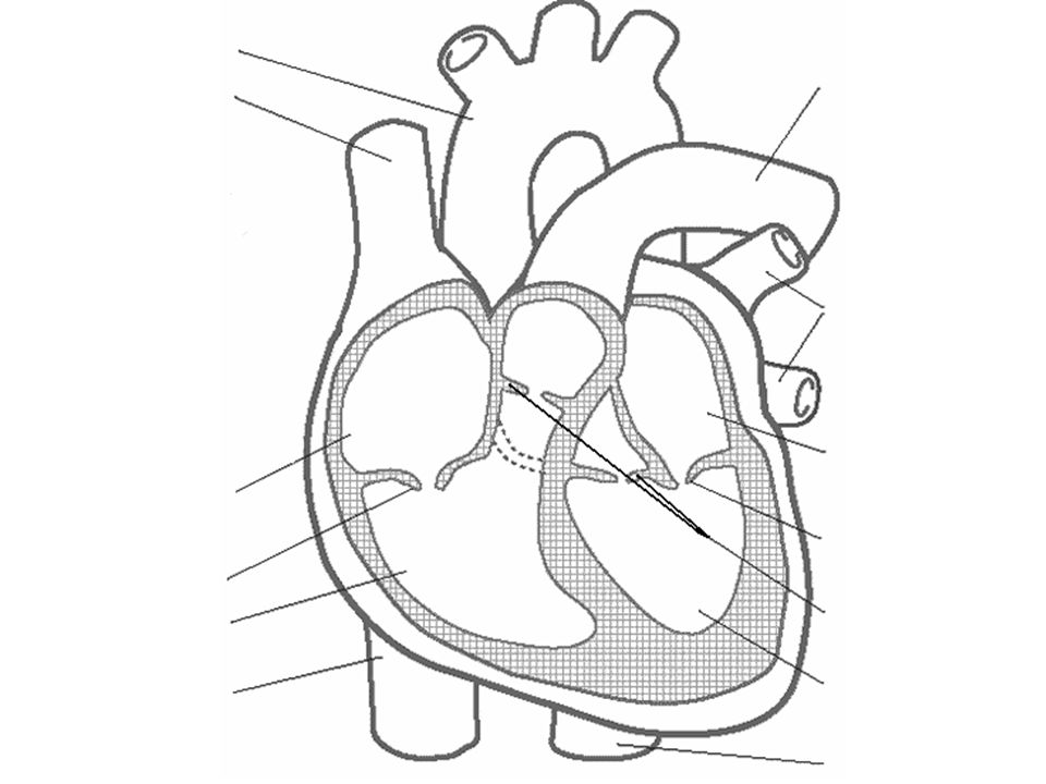
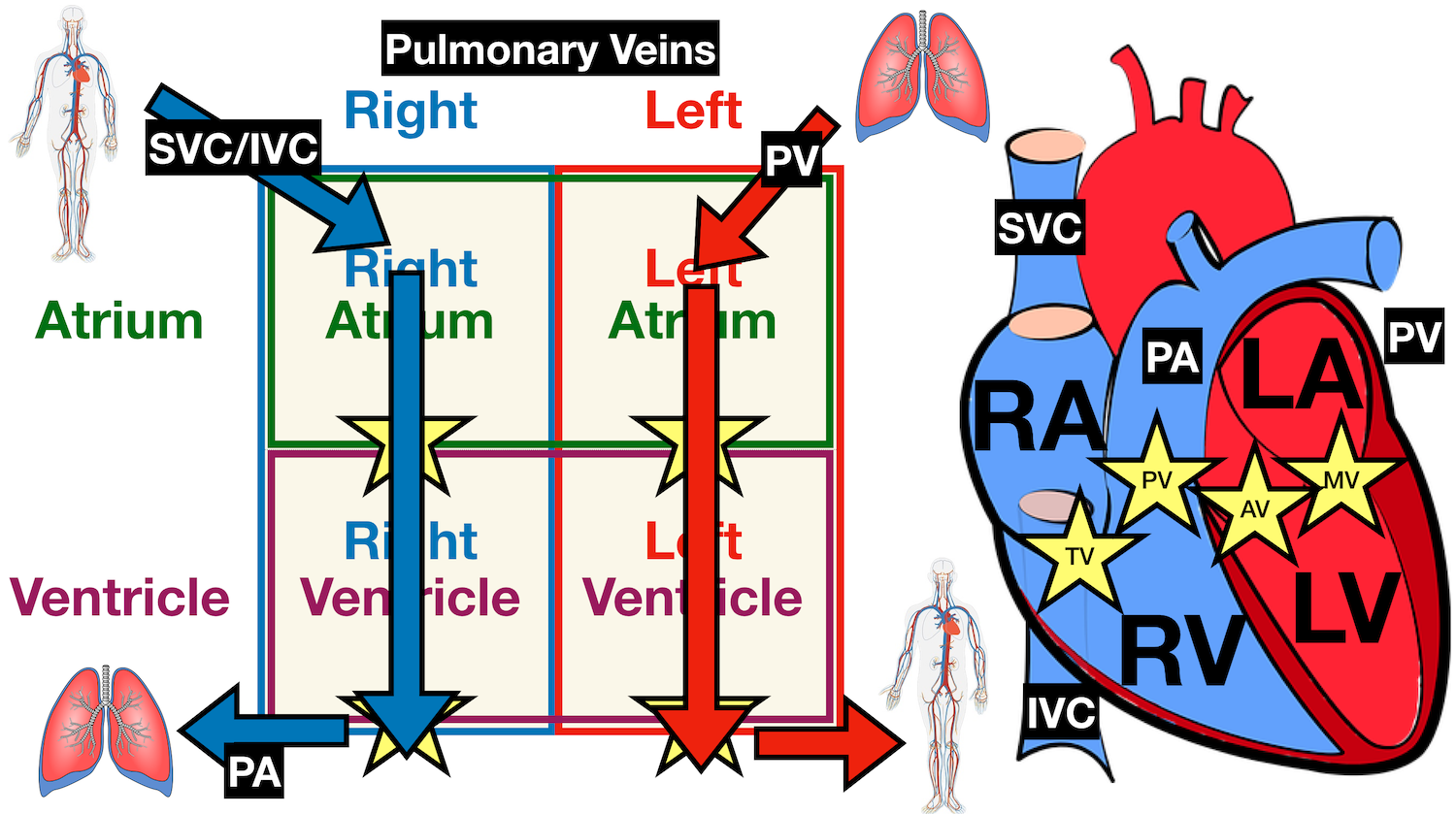
heart | Structure, Function, Diagram, Anatomy, & Facts A thin layer of tissue, the pericardium, covers the outside, and another layer, the endocardium, lines the inside. The heart cavity is divided down the middle into a right and a left heart, which in turn are subdivided into two chambers. The upper chamber is called an atrium (or auricle), and the lower chamber is called a ventricle. Heart anatomy: Structure, valves, coronary vessels | Kenhub Inside, the heart is divided into four heart chambers: two atria (right and left) and two ventricles (right and left).
Human Heart Models | Heart Anatomy Models | Vitality Medical The heart model with labels is hand-painted with vivid colors to illustrate the papillary muscles, heart valves, and adjacent structures. Sort By 4 Items Magnetic Heart Model, Life Size, 5 Parts $327.45 View Details Human Heart Model $450.66 - $566.36 View Details Classic Heart Model $81.03 View Details Magnetic Heart Model, Life Size, 5 Part G01
Heart structure and labels
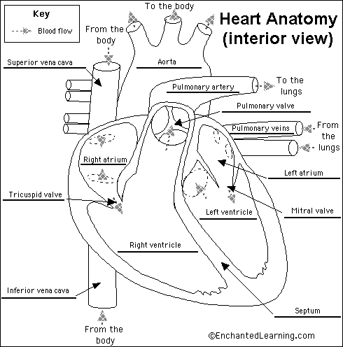
Human Heart Diagram Labeled | Science Trends List Of Heart Structures Heart Chambers Ventricles - The bottom two heart chambers. Atra - The upper two heart chambers. Wall Of The Heart Sinoatrial Node - A collection of tissue that releases electrical impulses and defines the rate of contraction for the heart. Atrioventricular Bundle - The fibers which transmit cardiac impulses. PDF Free Anatomy Coloring Page - North Carolina State University The ate.2S the heart With oxygen ate labeled with at'l Color these areas The areas o' the heart with less oxygen ate labeled with a color areas BLUE. ARTERY LEFT LUNG PULMONARY VEINS AORTA PULMONARY VEINS raGHT LUNG ATRIUM RIGHT VENTRICLE INFERIOR VFNACAVA LEFT LEFT VENTRICLE AORTA BODY Downloaded from azcoloring.com Label Heart Anatomy Diagram Printout - EnchantedLearning.com This cycle is then repeated. Every day, the heart pumps about 2,000 gallons (7,600 liters) of blood, beating about 100,000 times. Label the heart anatomy diagram below using the heart glossary. Note: On the diagram, the right side of the heart appears on the left side of the picture (and vice versa) because you are looking at the heart from the ...
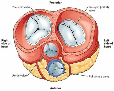
Heart structure and labels. Heart Diagram with Labels and Detailed Explanation - Collegedunia The heart is located under the ribcage, between the lungs and above the diaphragm. It weighs about 10.5 ounces and is cone shaped in structure. It consists of the following parts: Heart Detailed Diagram Heart - Chambers There are four chambers of the heart . The upper two chambers are the auricles and the lower two are called ventricles. Structure of the Heart | SEER Training - National Cancer Institute The human heart is a four-chambered muscular organ, shaped and sized roughly like a man's closed fist with two-thirds of the mass to the left of midline. The heart is enclosed in a pericardial sac that is lined with the parietal layers of a serous membrane. The visceral layer of the serous membrane forms the epicardium. Layers of the Heart Wall Structure and function of the heart - BBC Bitesize It is located in the middle of the chest and slightly towards the left. The heart is a large muscular pump and is divided into two halves - the right-hand side and the left-hand side. The... Heart: Anatomy and Function - Cleveland Clinic What are the parts of the heart's anatomy? The parts of your heart are like the parts of a house. Your heart has: Walls. Chambers (rooms). Valves (doors). Blood vessels (plumbing). Electrical conduction system (electricity). Heart walls Your heart walls are the muscles that contract (squeeze) and relax to send blood throughout your body.
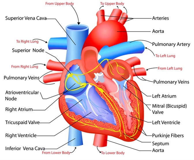
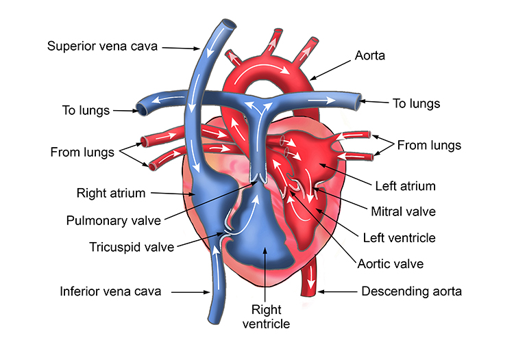
How to Draw the Internal Structure of the Heart (with Pictures) - wikiHow To draw the internal structure of a human heart, follow the steps below. Part 1 Finding a Diagram 1 To find a good diagram, go to Google Images, and type in "The Internal Structure of the Human Heart". Find an image that displays the entire heart, and click on it to enlarge it. 2 Find a piece of paper and something to draw with. 13+ Heart Diagram Templates - Sample, Example, Format Download Color Heart Diagram Sample Format Free Download. cdhb.health.nz This colored heart diagram is a graphic representation of the organ which can be used for presentations and videos about the subject of human heart. The picture is in a coloured format and is available for a free download. Free Download. Heart (Human Anatomy): Overview, Function & Structure | Biology The heart is a muscular organ that pumps blood throughout the body. It is located in the middle cavity of the chest, between the lungs. In most people, the heart is located on the left side of the chest, beneath the breastbone. The heart is composed of smooth muscle. It has four chambers which contract in a specific order, allowing the human ... How the Heart Works: Diagram, Anatomy, Blood Flow - MedicineNet Normal heart anatomy and physiology. Normal heart anatomy and physiology need the atria and ventricles to work sequentially, contracting and relaxing to pump blood out of the heart and then to let the chambers refill. When blood leaves each chamber of the heart, it passes through a valve that is designed to prevent the backflow of blood.
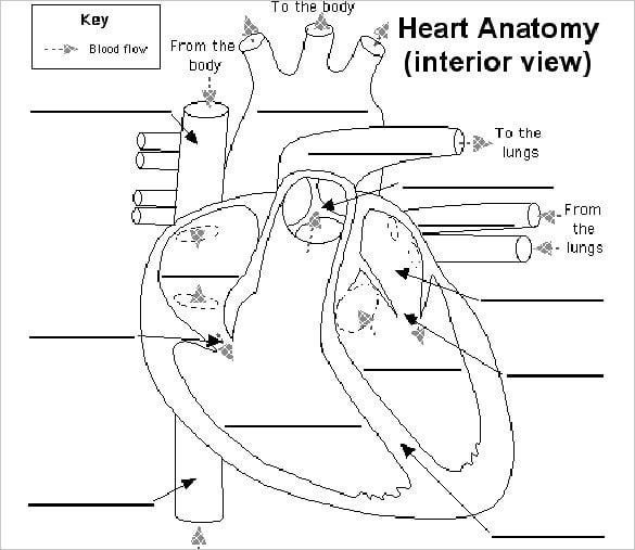
Heart Anatomy Labeling Game - PurposeGames.com This is an online quiz called Heart Anatomy Labeling Game There is a printable worksheet available for download here so you can take the quiz with pen and paper. Your Skills & Rank Total Points 0 Get started! Today's Rank -- 0 Today 's Points One of us! Game Points 19 You need to get 100% to score the 19 points available Actions Diagrams, quizzes and worksheets of the heart | Kenhub Worksheet showing unlabelled heart diagrams. Using our unlabeled heart diagrams, you can challenge yourself to identify the individual parts of the heart as indicated by the arrows and fill-in-the-blank spaces. This exercise will help you to identify your weak spots, so you'll know which heart structures you need to spend more time studying ... Heart Diagram with Labels and Detailed Explanation - BYJUS Diagram of Heart. The human heart is the most crucial organ of the human body. It pumps blood from the heart to different parts of the body and back to the heart. The most common heart attack symptoms or warning signs are chest pain, breathlessness, nausea, sweating etc. The diagram of heart is beneficial for Class 10 and 12 and is frequently ... Heart: illustrated anatomy - e-Anatomy - IMAIOS This interactive atlas of human heart anatomy is based on medical illustrations and cadaver photography. The user can show or hide the anatomical labels which provide a useful tool to create illustrations perfectly adapted for teaching. Anatomy of the heart: anatomical illustrations and structures, 3D model and photographs of dissection.
13+ Heart Diagram Templates – Sample, Example, Format ...
Heart Anatomy: size, location, coverings and layers : Anatomy & Physiology Heart Anatomy. The heart is around the size of a fist and weighs between 250-350 grams (less than a pound). Enclosed within the mediastinum, the medial cavity of the thorax, the heart extends obliquely from the second rib to the fifth intercostal space. It rests on the superior surface of the diaphragm, lies posterior to the sternum and ...
File:Diagram of the human heart (clean).svg - Wikimedia Commons
Structure of the Heart | The Franklin Institute The heart consists of four chambers: two atria on the top and two ventricles on the bottom. Looking at the Valentine's Day heart, the two rounded humps at the top are rounded like the top of a lower-case "a." The bottom is shaped like a "v." Feel it working What else is inside your heart?
(230).jpg)
Heart Labeling Quiz: How Much You Know About Heart Labeling ...
147 Heart Anatomy With Labels Premium High Res Photos - Getty Images Browse 147 heart anatomy with labels stock photos and images available, or start a new search to explore more stock photos and images. of 3. NEXT.

Heart Anatomy Vector Illustration Labeled Organ Stock Vector ...
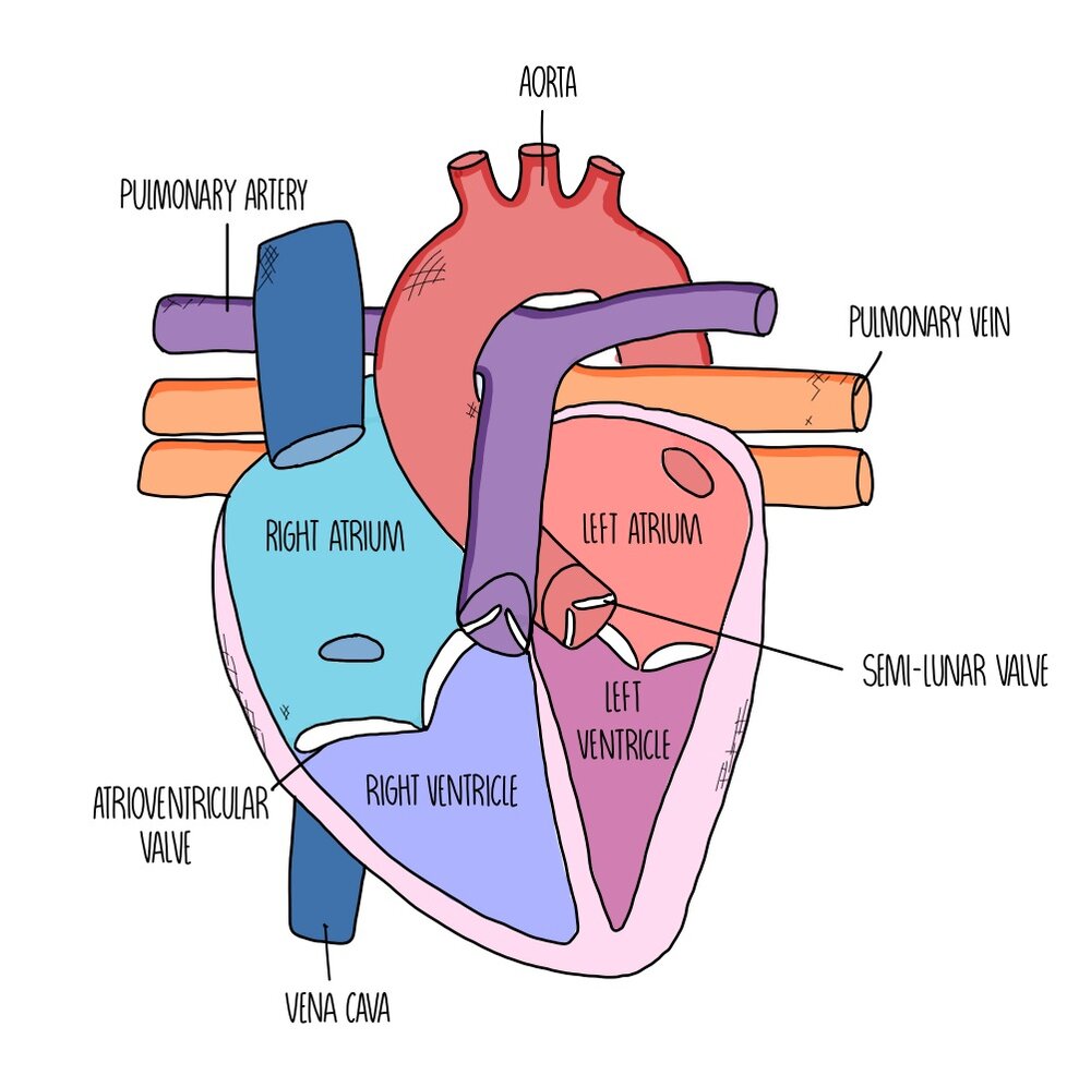
Label the heart — Science Learning Hub Label the heart Interactive Add to collection In this interactive, you can label parts of the human heart. Drag and drop the text labels onto the boxes next to the diagram. Selecting or hovering over a box will highlight each area in the diagram. pulmonary vein semilunar valve right ventricle right atrium vena cava left atrium pulmonary artery
Notes: Heart and Circulatory System
The Anatomy of the Heart, Its Structures, and Functions - ThoughtCo The heart is the organ that helps supply blood and oxygen to all parts of the body. It is divided by a partition (or septum) into two halves. The halves are, in turn, divided into four chambers. The heart is situated within the chest cavity and surrounded by a fluid-filled sac called the pericardium. This amazing muscle produces electrical ...

draw and label the structure of a human heart - Brainly.in
Labelling the heart — Science Learning Hub The heart is a muscular organ that pumps blood through the blood vessels of the circulatory system. Blood transports oxygen and nutrients to the body. It is also involved in the removal of metabolic wastes. In this activity, students use online and paper resources to identify and label the main parts of the heart.
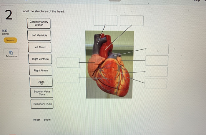
Solved Label the structures of the heart. Coronary Artery ...
Human Heart (Anatomy): Diagram, Function, Chambers, Location in ... - WebMD Chambers of the Heart The heart is a muscular organ about the size of a fist, located just behind and slightly left of the breastbone. The heart pumps blood through the network of arteries and...
The Structure of the Heart Learning Objectives: Label the ...
Structure and Function of the Heart - News-Medical.net Structure of the heart The heart wall is composed of three layers, including the outer epicardium (thin layer), middle myocardium (thick layer), and innermost endocardium (thin layer). The...
Anatomy of the Human Heart - Physiopedia
20+ Simple heart diagram | Simple heart diagram labeled - Pinterest Dec 23, 2021 - Simple heart diagram | Simple heart diagram labeled | Human heart diagram.We provide you a simple heart diagram to draw and learn. Simple heart diagram labeled with accurate labels. Most frequent question in exam to draw human heart diagram with labels. You can learn diagram of heart with labels and easy simple heart anatomy with heart structure. Learn to draw Simple heart ...

4,122 Human Heart Diagram Stock Photos, Pictures & Royalty ...
Heart Labels - Printable or Custom Printed Stickers | Avery.com Use our free specialty shape label templates to easily personalize your heart labels online. Customize one of our free designs or upload your own graphics and then choose the printing option that works best for you. Order your blank heart labels or custom printed heart labels and stickers online and get free shipping on orders of $50 more.

Colorful Hand Drawn Illustration Of Human Heart Anatomy Stock ...
Heart Labeling Quiz: How Much You Know About Heart Labeling? Here is a Heart labeling quiz for you. The human heart is a vital organ for every human. The more healthy your heart is, the longer the chances you have of surviving, so you better take care of it. Take the following quiz to know how much you know about your heart. Questions and Answers 1. What is #1? 2. What is #2? 3. What is #3? 4. What is #4?
Heart Structure and Cardiac Cycle (A Level) — the science hive
Human Heart - Diagram and Anatomy of the Heart - Innerbody Because the heart points to the left, about 2/3 of the heart's mass is found on the left side of the body and the other 1/3 is on the right. Anatomy of the Heart Pericardium. The heart sits within a fluid-filled cavity called the pericardial cavity. The walls and lining of the pericardial cavity are a special membrane known as the pericardium.

File:Heart diagram-en.svg - Wikimedia Commons
Label Heart Anatomy Diagram Printout - EnchantedLearning.com This cycle is then repeated. Every day, the heart pumps about 2,000 gallons (7,600 liters) of blood, beating about 100,000 times. Label the heart anatomy diagram below using the heart glossary. Note: On the diagram, the right side of the heart appears on the left side of the picture (and vice versa) because you are looking at the heart from the ...

Heart Anatomy: Labeled Diagram, Structures, Blood Flow ...
PDF Free Anatomy Coloring Page - North Carolina State University The ate.2S the heart With oxygen ate labeled with at'l Color these areas The areas o' the heart with less oxygen ate labeled with a color areas BLUE. ARTERY LEFT LUNG PULMONARY VEINS AORTA PULMONARY VEINS raGHT LUNG ATRIUM RIGHT VENTRICLE INFERIOR VFNACAVA LEFT LEFT VENTRICLE AORTA BODY Downloaded from azcoloring.com
Structure of the heart | Quiz
Human Heart Diagram Labeled | Science Trends List Of Heart Structures Heart Chambers Ventricles - The bottom two heart chambers. Atra - The upper two heart chambers. Wall Of The Heart Sinoatrial Node - A collection of tissue that releases electrical impulses and defines the rate of contraction for the heart. Atrioventricular Bundle - The fibers which transmit cardiac impulses.

Activity
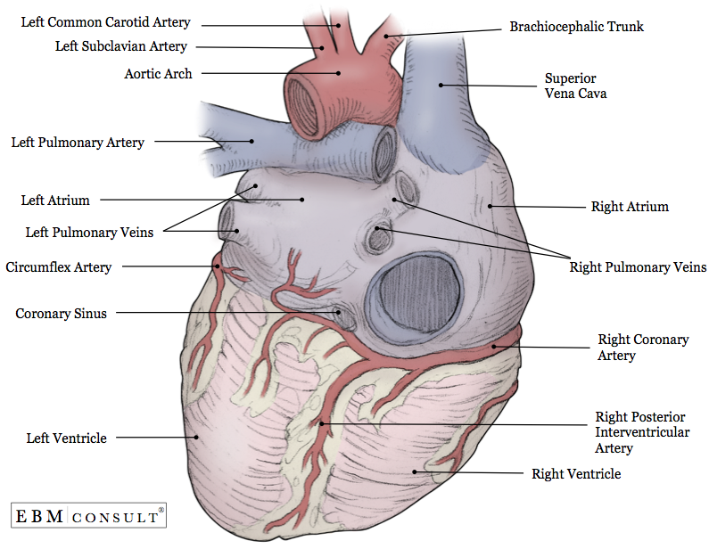
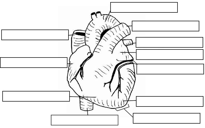
Anatomy: Heart (External)
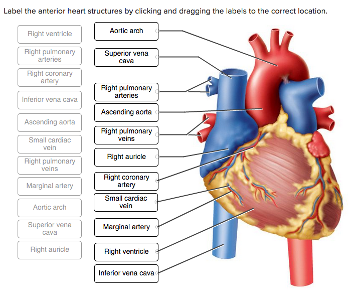
Solved Label the anterior heart structures by clicking and ...
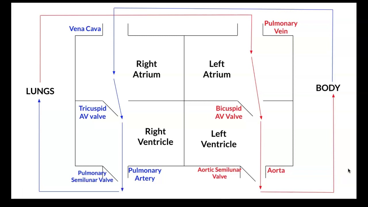
Box Diagram, Labels of Heart, and Blood Flow through Heart
Heart Anatomy Glossary Printout - EnchantedLearning.com
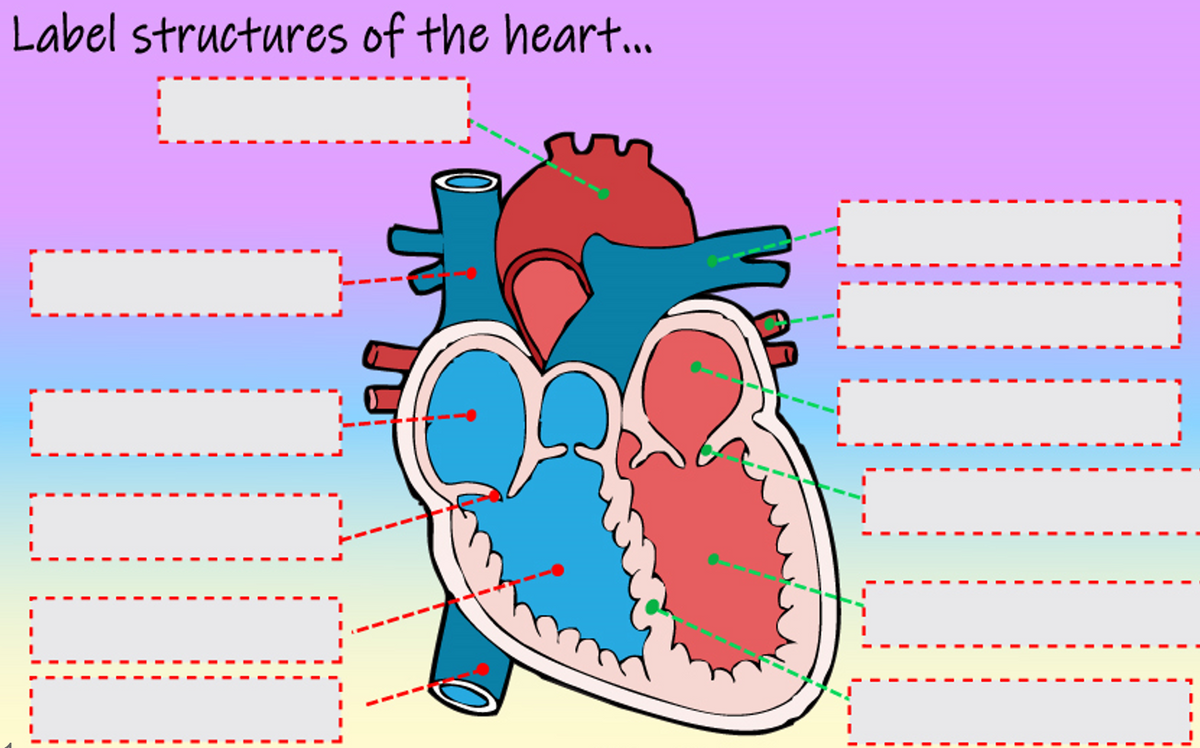
Answered: Label structures of the heart... | bartleby

Pin on Paramedic Study Guide

4,122 Human Heart Diagram Stock Photos, Pictures & Royalty ...

Label parts of the heart worksheet
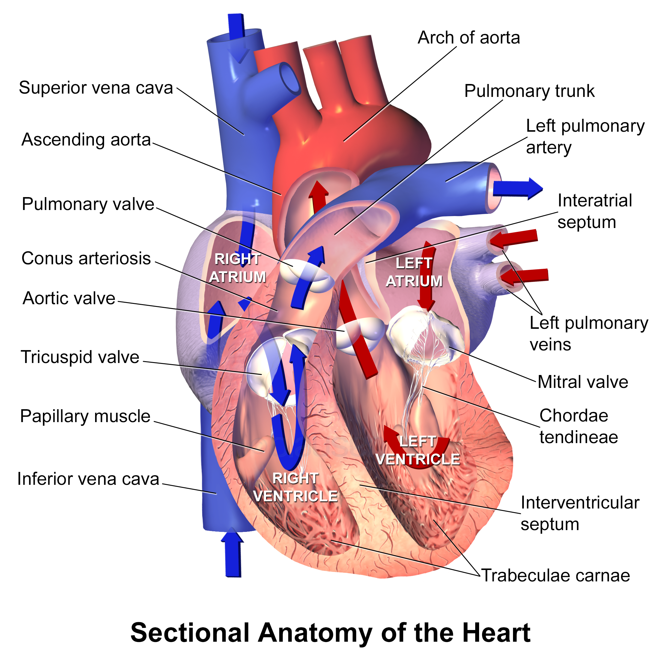
Blausen 0457 - Sectional anatomy of the heart - English ...

Sketch the internal structure of human heart. Label all the ...
Structure and Function of the Heart
Revision notes of heart structure and labelled diagram
Diagrams, quizzes and worksheets of the heart | Kenhub

4,122 Human Heart Diagram Stock Photos, Pictures & Royalty ...
Heart Anatomy: Labeled Diagram, Structures, Blood Flow ...

Solved: Label the following structures of the heart by ...
The Heart - Label and describe by Leah Mulvey | Teachers Pay ...
Heart Diagram with Labels and Detailed Explanation

Simple heart diagram | Simple heart diagram labeled | Human ...

Label this: posterior surface heart structures Diagram | Quizlet
Free Unlabelled Diagram Of The Heart, Download Free ...
Heart Anatomy: Labeled Diagram, Structures, Blood Flow ...
Label the heart — Science Learning Hub
Learn the Anatomy of the Heart
Post a Comment for "40 heart structure and labels"