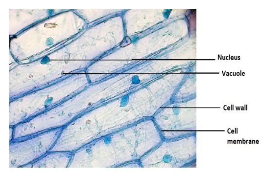
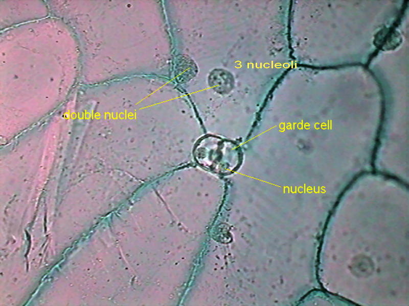
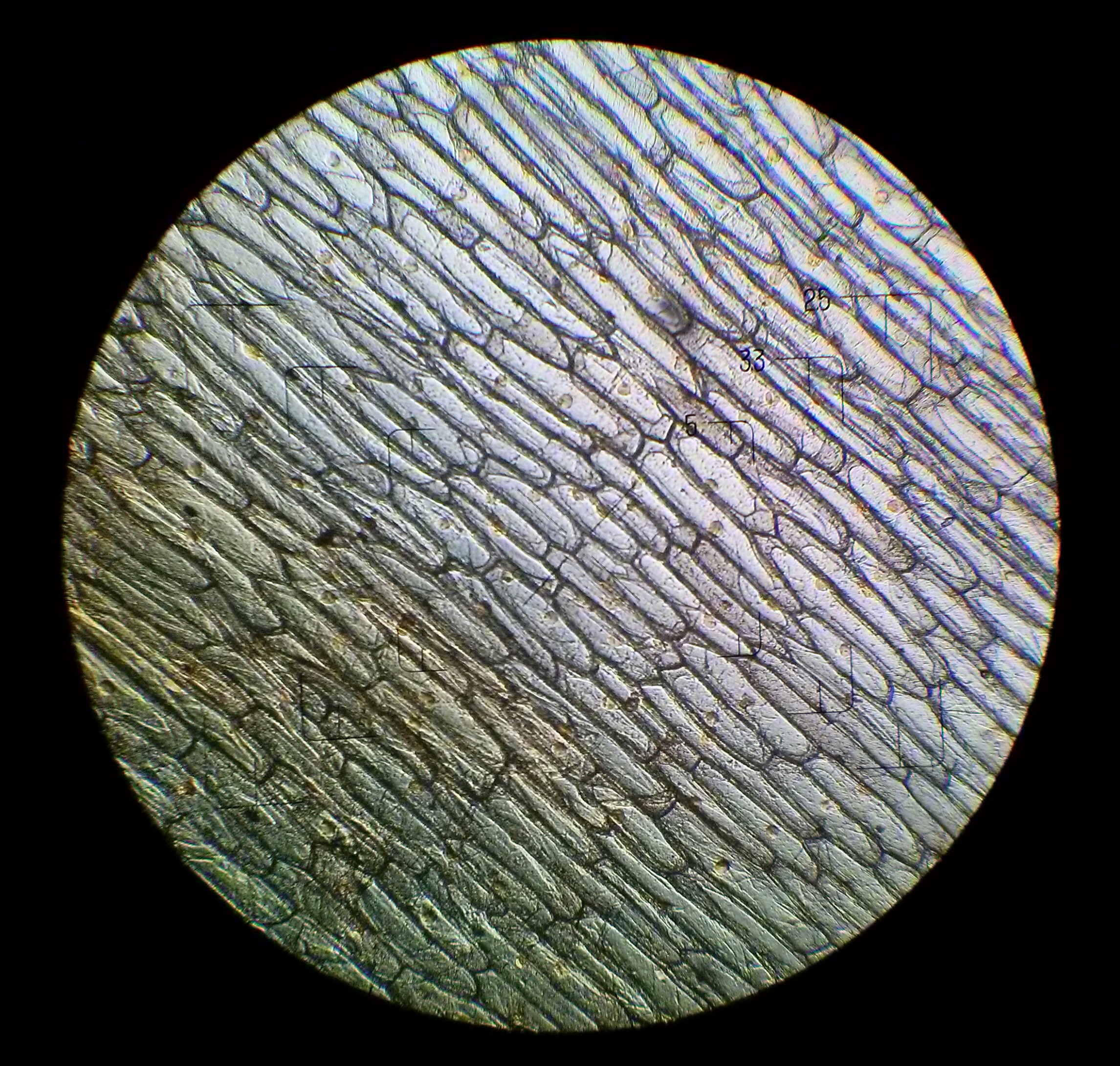
42 onion cells under microscope with labels
Satellite News and latest stories | The Jerusalem Post Mar 08, 2022 · The Jerusalem Post Customer Service Center can be contacted with any questions or requests: Telephone: *2421 * Extension 4 Jerusalem Post or 03-7619056 Fax: 03-5613699 E-mail: [email protected ... Scanning electron microscope - Wikipedia History. An account of the early history of scanning electron microscopy has been presented by McMullan. Although Max Knoll produced a photo with a 50 mm object-field-width showing channeling contrast by the use of an electron beam scanner, it was Manfred von Ardenne who in 1937 invented a microscope with high resolution by scanning a very small raster with a demagnified and finely focused ...
Microscopy Practical (Onion Cells) | Teaching Resources pptx, 12.37 MB. pdf, 223.81 KB. docx, 1.09 MB. Presentation and practical handout for observing onion cells under a light microscope for teaching and revision. A step by step visual guide for all abilities. Can be used as a distance based learning tool during local covid lockdown and in classes where practicals are on-hold due to coronavirus.

Onion cells under microscope with labels
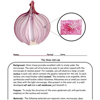
Nano based drug delivery systems: recent developments and ... Sep 19, 2018 · Nanomedicine and nano delivery systems are a relatively new but rapidly developing science where materials in the nanoscale range are employed to serve as means of diagnostic tools or to deliver therapeutic agents to specific targeted sites in a controlled manner. Nanotechnology offers multiple benefits in treating chronic human diseases by site-specific, and target-oriented delivery of ... Microscope Cell Lab: Cheek, Onion, Zebrina | SchoolWorkHelper The onion epidermis cell is the only cell that has a cell wall. In addition, it is the only cell that has a chloroplast, where photosynthesis can happen. The cheek epithelium cell is the only one that has centrioles, the barrel-shaped organelle that is responsible for helping organize chromosomes during cell division. The Omnivore's Dilemma: A Natural History of Four Meals ... "Outstanding . . . a wide-ranging invitation to think through the moral ramifications of our eating habits." — The New Yorker One of the New York Times Book Review's Ten Best Books of the Year and Winner of the James Beard Award Author of How to Change Your Mind and the #1 New York Times Bestseller In Defense of Food and Food Rules What should we have for dinner?
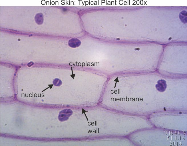
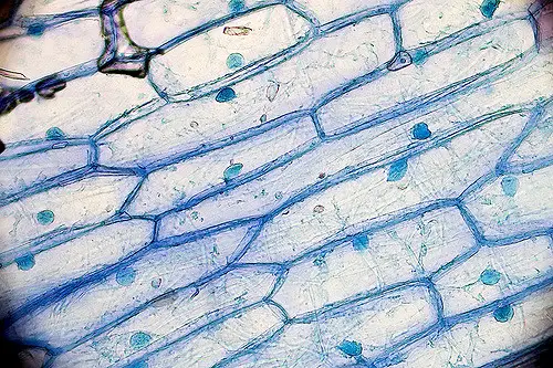
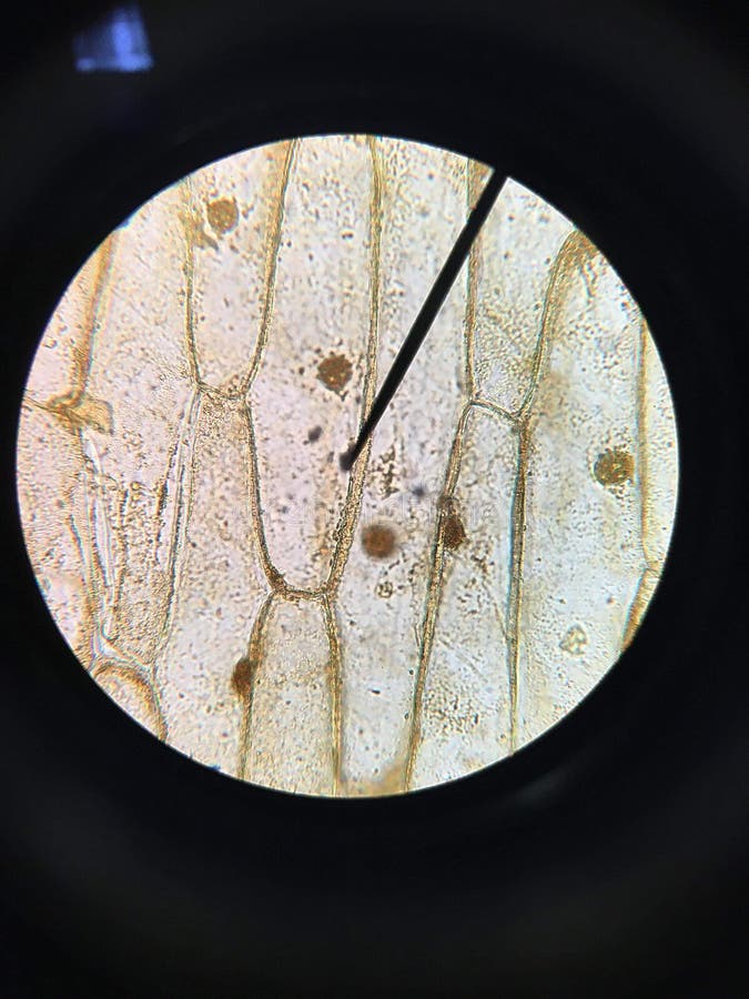
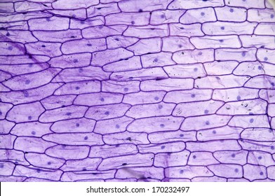
Onion cells under microscope with labels. What organelles are in an onion cell? - Biology Stack Exchange You cannot see most of these as they appear translucent as well as being too small to see under the light microscope. You need an electron microscope to view these. Note: chloroplasts are not present in an onion cell as it is not a photosynthesising cell. This is a typical onion cell slide with labels: Science — Biology – Easy Peasy All-in-One Homeschool See if you can see the parts of a bulb in an onion or garlic clove. Finish reading the rest of the case. Keep clicking next. Level 5-8. Read “What is a bulb?” and follow the directions. Cut open an onion or garlic clove. These are bulbs. Plants can be grown from these. Do you ever see green leaves coming out of your onions or garlic? Onion Plant Cell Under Microscope Labeled - Ismael Dauila Explore diffusion/osmosis by looking at onion cells under the microscope. It is used for treating a parasite disease called ich (ichthyophthirius multifiliis; Label the cell wall and chloroplasts. Students will observe plant cells using a light microscope. Lesson 3: Onion Dissection & "Look at the Plant Cells" Preparing onion cells slide for a microscope Peel the brown skin away from the outside of the onion. Take one layer of the onion flesh and carefully cut out a piece. On the inside of this piece is a thin sheet of the membrane. Use tweezers or dissection needle to peel the membrane away.
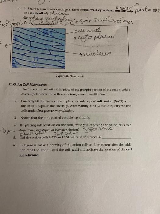
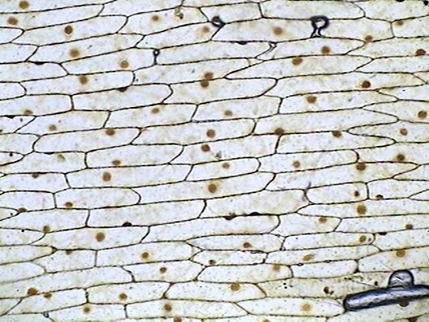
Onion Peel Cell Experiment - Biology Reader Compound microscope; Theory. An onion is a multicellular plant. The presence of a rigid cell wall and a large vacuole is a characteristic feature of a plant cell. Thus, onion being a plant, comprises features common to plant cells. Like plant cells, onion cells consist of a cell wall and cell membrane surrounding the cytoplasm, nucleus and a ... Looking at the Structure of Cells in the Microscope Both types of light microscopy are widely used to visualize living cells. Figure 9-7 Two ways to obtain contrast in light microscopy. (A) The stained portions of the cell reduce the amplitude of light waves of particular wavelengths passing through them. A colored image of the cell is thereby obtained that is visible in the ordinary way. (more...) DOC Plant and Animal Cells Microscope Lab - hillsboro.k12.oh.us Make a drawing of one onion cell, labeling all of its parts as you observe them. (At minimum you should observe the nucleus, cell wall, and cytoplasm.) Cheek cells 1. To view cheek cells, gently scrape the inside lining of your cheek with a toothpick. DO NOT GOUGE THE INSIDE OF YOUR CHEEK! (We will observe blood cells in a future lab!!) 2. Onion Peels Observed Under the Microscope | Confirmation Point Onion Peels Observed Under the Microscope Cells present in onion peel can be observed under microscope. For this onion peels are first isolated. For this experiment outer most scale of the onion is removed and is cut into four equal halves. It is a monocot plant. Then with the help of a pairs of forcep the scale of onion is peeled out.
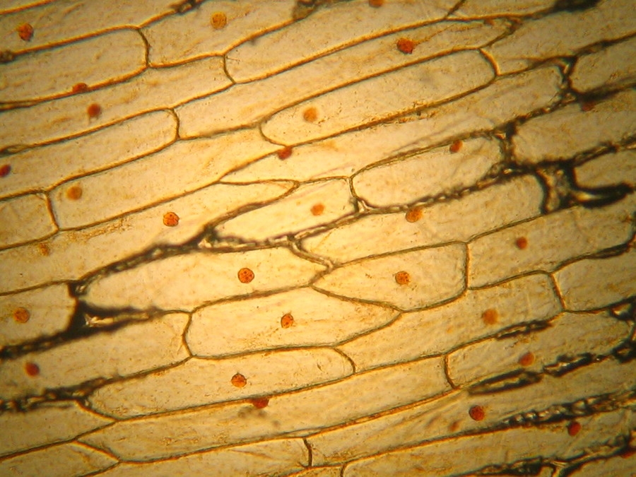
Onion peel under microscope | AQuriousMind Prepare the onion peel slide Use water to clean the slides and cover slips. Use the knife to cut a small square section of the onion. Remove the first layer (its a leaf!) and then further cut it into two or four parts. The smaller the specimen the better. Use the tweezer to pull out one thin peel of the onion. This peel is exactly one cell thick. Observing Onion Cells Under The Microscope » Microscope Club Afterwards, carefully mount the prepared and stained onion cell slide onto the microscope stage. Make sure that the cover slip is perfectly aligned with the microscope slide, and that any excess stain has been wiped off. Secure the slide on the stage using the stage clips. Onion Cells Under a Microscope - Requirements/Preparation/Observation Add a drop of iodine solution on the onion membrane (or methylene blue) Gently lay a microscopic cover slip on the membrane and press it down gently using a needle to remove air bubbles. Touch a blotting paper on one side of the slide to drain excess iodine/water solution, Place the slide on the microscope stage under low power to observe. Onion Root Tip Mitosis - Stages, Experiment and Results - MicroscopeMaster · Place a cap/lid onto the vial (ensure that the cap/lid has a pinprick hole) and place the vial in the water bath (at 55 degrees C) for about 5 minutes - This enhances the staining process · Using the forceps, remove the root tips from the vial of stain and place them onto a clean microscope glass slide
Cell structure Learning Intention: - ppt video online download
The Biology Project The Biology Project, an interactive online resource for learning biology developed at The University of Arizona. The Biology Project is fun, richly illustrated, and tested on 1000s of students. It has been designed for biology students at the college and high school level, but is useful for medical students, physicians, science writers, and all types of interested people.
Solved] Fig. 2.7. Stained cells of fleshy onion (Allium cepa ...
Under the Micrsocope: Onion Cell (100x - 400x) - YouTube In this "experiment" we will see onion cells under the microscope.For the experiment you will only need onion, dropper and the microscope (container and tool...

How to Observe Onion Cells under a Microscope - Blog, She Wrote
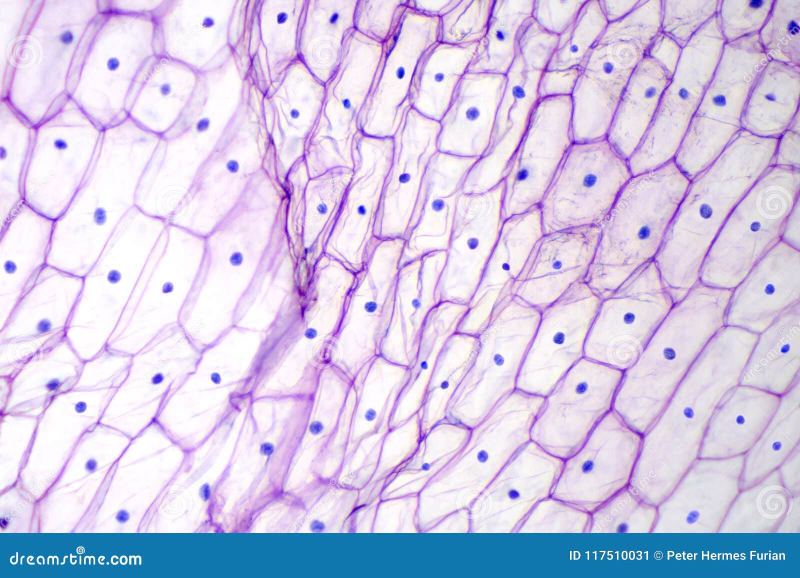
Cambridge IGCSE Biology Coursebook (third edition) - Issuu Jun 09, 2014 · If you colour or stain the cells, they are quite easy to see using a light microscope (see Figure 2.6 and Figure 2.11). 1 Using a section lifter, gently rub off a little of the lining from the ...

Onion Skin Epidermal Cells: How to Prepare a Wet Mount Microscope Slide
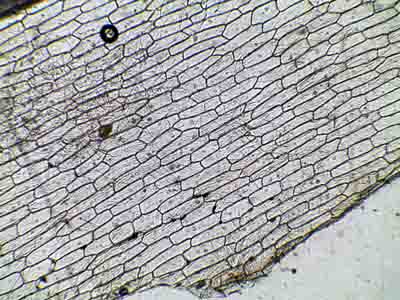
Onion cells microscope Stock Photos and Images - Alamy RM2AM97C0-Onion skin cells under the microscope, horizontal field of view is about 0.61 mm RFHWA476-Onion epidermis with large cells under light microscope. Clear epidermal cells of an onion, Allium cepa, in a single layer. RM2DF6FFJ-Onion epidermis (Allium cepa) showing cells and nucleus. Optical microscope X200.

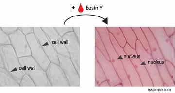
Why is iodine stain used on onion cells? | Iodine, Cell, Stain
DOC The Onion Cell Lab - chsd.us Place the single layer of onion cell epithelium on a glass slide. Make sure that you do not fold it over or wrinkle it. Place a drop of iodine stain on your onion tissue. Put the cover slip on the stained tissue and gently tap out any air bubbles. Observe the cells under 4x, 10x, and 40x with the diaphragm wide open.
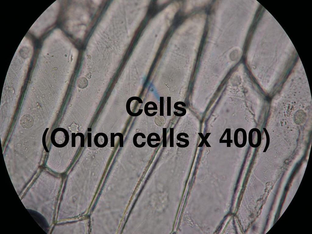
Cells (Onion cells x 400). - ppt download
A Level Biology For OCR A (PDFDrive) | PDF - Scribd Light microscopy started the science of cell biology, but it has limitations. In the middle of the 20th century a new invention, the electron microscope, revolutionised the study of cells and enabled biologists to see deep inside structures that were invisible under a light microscope.

Structure and Function of Cells (Learn) : Biology : Class 8 ...
Onion cells under the microscope: 40X - 100X - 400X - YouTube under the #microscope: 40X - 100X - 400X
Onion skin 200x - Dissection Connection
Observing Cork Cells Under The Microscope » Microscope Club Place the cork dust on the microscope slide with a drop of water, then add another water droplet on top of the cork sample. Cover the prepared slide with a cover slip. Method 2 Alternatively, slice thin cork slices, making sure that ample light can pass through the slice, allowing you to see the cell layout and the individual cells.
Onion Cell Microscope Lab
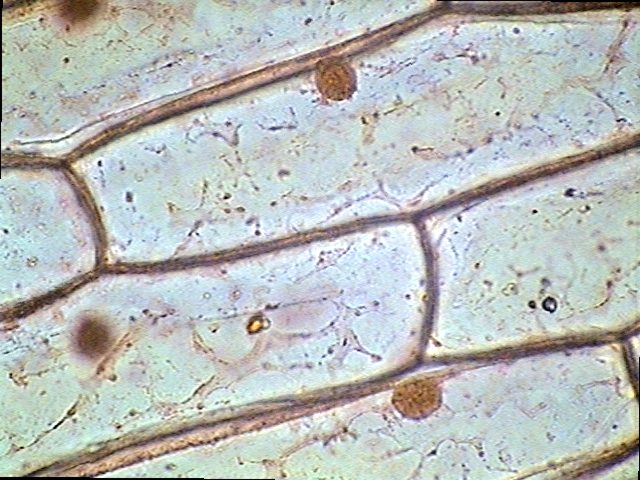
The Cell Structure of an Onion | Sciencing Onion cells are among the most common choices for cell studies in early biology classes. Easily obtained, inexpensive, they offer samples with no difficult technique required. The thin layer of skin found on the inside of an onion scale (one layer of onion) lifts off without effort and can be wet mounted on a slide with no need for extreme skill.

Tonicity-Onion cell lab | Miranda's AP Biology Blog
Onion Cell Lab Report.docx - Onion Cell Lab Report By Onion Cell Lab Report By : Nawaf Almalki Introduction: Many things that are viewed using a microscope, particularly cells, can appear quite transparent under the microscope. The internal parts of the cells, the organelles, are so transparent that they are often difficult to see. Biologists have developed a number of stains that help them see the cells and their organelles by adding color to ...

onion cells - Google Images | Ilustrações, Felipão, Estampas
Onion Cells Under Microscope With Labels - Realtec Find and download Onion Cells Under Microscope With Labels image, wallpaper and background for your Iphone, Android or PC Desktop. Realtec have about 34 image published on this page. onion microscope under cells cepa allium slide footage shutterstock background royalty Pin It Share Download
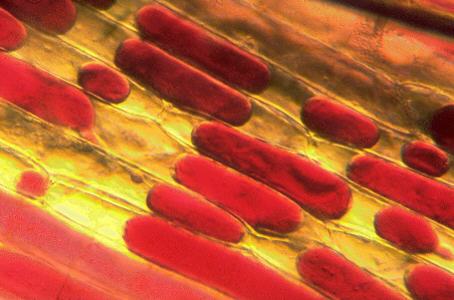
Mic-UK: MICROSCOPY UK / MICSCAPE - Onion epidermis, plasmolysis
Onion Root Mitosis - Microscopy-UK Onions have larger chromosomes than most plants and stain dark. The chromosomes are easily observed through a compound light microscope. The cells pictured below are located in the apical meristem of the onion root. The apical meristem is an area of a plant where cell division takes place at a rapid rate. Phases of plant cells division:
General Biology Microscopic Specimen Images & Photographs
The Omnivore's Dilemma: A Natural History of Four Meals ... "Outstanding . . . a wide-ranging invitation to think through the moral ramifications of our eating habits." — The New Yorker One of the New York Times Book Review's Ten Best Books of the Year and Winner of the James Beard Award Author of How to Change Your Mind and the #1 New York Times Bestseller In Defense of Food and Food Rules What should we have for dinner?

Cells Under A Microscope by Jaimarie Nelson
Microscope Cell Lab: Cheek, Onion, Zebrina | SchoolWorkHelper The onion epidermis cell is the only cell that has a cell wall. In addition, it is the only cell that has a chloroplast, where photosynthesis can happen. The cheek epithelium cell is the only one that has centrioles, the barrel-shaped organelle that is responsible for helping organize chromosomes during cell division.
Solved 4. Using the low power objective, view several potato ...
Nano based drug delivery systems: recent developments and ... Sep 19, 2018 · Nanomedicine and nano delivery systems are a relatively new but rapidly developing science where materials in the nanoscale range are employed to serve as means of diagnostic tools or to deliver therapeutic agents to specific targeted sites in a controlled manner. Nanotechnology offers multiple benefits in treating chronic human diseases by site-specific, and target-oriented delivery of ...

Identifying the Structures That Can Be Observed under a Microscope

Epidermal onion cells under a microscope. Plant cells appear ...

Onion Epidermis
Plant Cell Lab - Onion and Elodea

Under The Microscope Onion Cells Stock Photo, Picture And ...

1.1 Cell structure | Cells as the basic units of life | Siyavula
Onion Cells Under a Microscope - Requirements/Preparation ...
Onion Cell Under Microscope 40X Stock Image - Image of cell ...
AIM To prepare stained temporary mount of onion peel class 7 ...

onion cells under microscope at over 300X !

Microscope Lc - Lessons - Blendspace
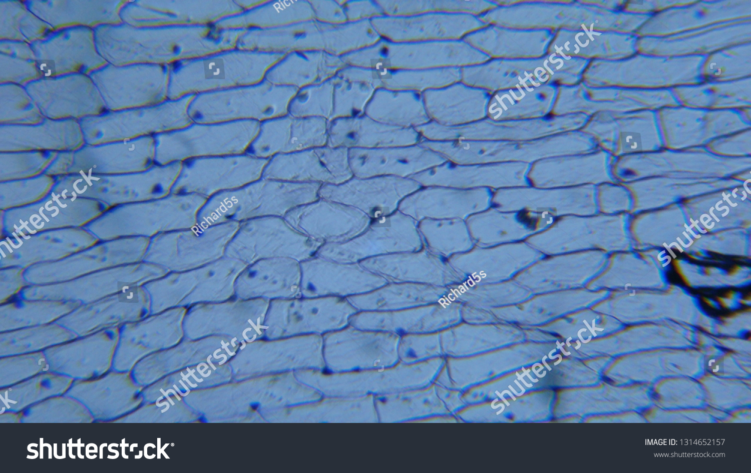
Onion Cells Under Microscope Stock Photo 1314652157 ...
Laboratory activity 3 (post lab)
167,485 Cell Wall Images, Stock Photos & Vectors | Shutterstock

Onion Cells at 400X Magnification
Mic-UK: Homage to the onion skin

Onion cells
Mic-UK: The inner epidermis of the onion bulb's cataphylls ...
Mic-UK: How many onion skins are there?
File:Onion cells under the light microscope.jpg - Wikimedia ...
Onion Epidermis with Large Cells Under Microscope Stock Image ...
Lesson 3: Onion Dissection & “Look at the Plant Cells” - Rs ...

microscopy how a microscope works magnification calculations ...

Lab #1 microscope structure & function
Biology

Onion Cell Microscope Slide Experiment - YouTube

School Science/How to prepare an onion cell slide - Wikibooks ...
Post a Comment for "42 onion cells under microscope with labels"